Diseases of the Australian Freshwater Fish Silver Perch (Bidyanus bidyanus) - Part 6
STREPTOCOCCOSIS
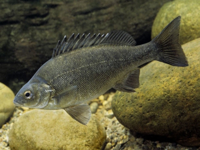
The bacterium, Streptococcus iniae, causes streptococcosis in silver perch. The disease has been rarely reported, although losses in the few outbreaks were significant. Streptococcus spp. are responsible for disease in many cultured fish worldwide, including rainbow trout, eels, channel catfish, tilapia, yellowtail and striped bass. The disease typically takes the form of septicaemia, and clinically fish may exhibit exophthalmia (‘popeye’) (Fig. 80) or fluid in the abdominal cavity and intestine (ascites).
Transmission of Streptococcus spp. is thought to be mainly by contact and is likely to be enhanced by injury to epithelium or by stressful environmental conditions. Natural epizootics have been recorded in populations of wild fish. Silver perch reared in RAS were infected following fluctuating water quality (18 to 11°C over 12 h); however, other factors may have contributed to the epizootic.
Pathogen
Grampositive, ovoid to elongate cocci; immotile; single or paired but rarely form chains in infected fish; β-haemolytic; many species can grow anaerobically, in a wide temperature range (10–45°C).
Signs
Chronic fish mortality/morbidity
Fish darkly pigmented
Exophthalmia and abdominal distension
Lethargy; fish near the surface and at pond edges
Loss of appetite and nervous behaviour
Diffuse haemorrhages around operculum, base of fins and skin, and inside of abdominal cavity (Fig. 81) Survivors may develop spinal deformity (scoliosis)


Diagnosis
Bacterial isolation from a variety of organs (spleen, liver, brain, eye and kidney); positive, bacterial culture produces dull, yellowish/grey, slightly raised and rounded, 1 to 2 mm colonies at 48 hours on blood agar media.
Treatment
Tanks:
selection of antibiotic should be based on laboratory sensitivity testing as S. iniae has variable sensitivity. Whilst awaiting culture results fish can be started on oxytetracycline, 20 mg/L active ingredient, 7 days at 20–30°C or 10 days at <20°C; maintain low light levels, good aeration; water exchange to ‘dilute’ bacterial load and retreat.
Ponds:
Oral administration via feed:
Oxytetracycline, 75 mg/kg fish for 10 days.
Prevention
Avoid stress due to poor water quality, overcrowding, overfeeding, feeding old feed or unnecessary handling during critical periods; infected fish or carcasses should be removed; dry and lime ponds regularly; disinfect tanks, floors and equipment, sodium hypochlorite (500 mg/L for 2 mins), benzalkonium chloride (1,000 mg/L for 20 mins).
MYCOBACTERIOSIS
Mycobacteriosis is a chronic bacterial disease that can be persistent in recirculating systems such as those used for barramundi, Murray cod and aquarium fish (Figs. 82 and 83). The disease has been recorded in over 150 species of marine and freshwater fish worldwide. The common strains most frequently isolated include Mycobacterium marinum, M. chelonae and M. fortuitum although other environmental strains have been isolated from infected fish; all have a worldwide distribution. Mycobacteriosis has been recorded in pondreared silver perch; however, the disease isn’t a significant threat to the industry at present. Large silver perch (500 g) with mycobacteriosis exhibited typical white nodules (granulomas) on the viscera, particularly the spleen; approximately 20% mortality was recorded over a 2 month period. The infection propagated in the pond with prevalence increasing from approximately 20% to 60% over 8 weeks. Mycobacterium spp. can infect humans (zoonotic); however, the risk is low; gloves should be worn when handling fish suspected of having any bacterial diseases. Cuts, spike wounds and abrasions should be rapidly cleaned and treated with a topical disinfectant to minimise the risk of infection.

Pathogen
Straight or slightly curved, immotile rods, 0.4 × 1.0 to 4.0 µm long; gram positive, acid fast; often staining unevenly; are slow to grow (up to 10 weeks required) and can be difficult to culture.
Signs
Chronic fish mortality/morbidity
Irregular swimming, fish at pond edges and surface
Loss of appetite
Emaciation and poor growth
Abnormal dark colour of skin
Shallow to deep ulcers
Fin erosion
Granulomas in viscera (kidney, spleen, heart), 1 to 4 mm diameter (Fig. 84)
Exophthalmia and abdominal swelling (ascites)
Diagnosis
Laboratory culture of bacterium; diagnosed by acidfast and gram staining (ZiehlNielson stained) of smears; histological sections of tissue granulomas (Fig. 85); microscopic examination, 200×, of wet, squashed mounts of kidney and spleen tissue for granulomas; the latter may include peripheral, lighter coloured, inflammatory cells.

Treatment
Once established, can be difficult to control or eradicate.
Tanks:
remove infected fish; clean contaminated tanks, pipes and equipment; disinfect equipment using sodium hypochlorite (10,000 mg/L chlorine reported level required to kill mycobacteria).
Ponds:
remove infected batch of fish; dry, desilt and lime pond.
Prevention
Mycobacterium spp. can survive long periods in the environment and many strains are natural soil organisms; transmission probably via the shedding of bacteria from infected skin ulcers and ‘bullying’ of infected fish; dry ponds between crops; lime ponds with CaO or Ca(OH)2; disinfect tanks and equipment; utilise UV and/or ozone units; remove infected and dead fish; maintain good water quality and husbandry practices; minimise damage to fish to prevent superficial infections.
AEROMONAD DERMATITIS (Motile Aeromonad Infection, MAI)
Aeromonad infections are one of the most common bacterial diseases f freshwater fish. Many species are susceptible, including silver perch.
Aeromonad bacteria are ubiquitous and found in most freshwater ponds, rivers and bottom mud, utilising organic material as a nutrient source. The main species infecting fish are Aeromonas hydrophila, A. sobria, and A. caviae. The disease in silver perch is observed postharvest during late spring and summer (causing ‘summer spots’) particularly when stocking densities in purging systems are increased. Clinical signs begin as depigmentation of areas of the epidermis on the caudal peduncle, top of the head, flanks and base of the pectoral fins (Fig. 86). If left untreated, ‘spots’ can develop into more advanced, red, necrotic skin lesions.

Mild lesions can progress rapidly, with significant scale loss and haemorrhaging around the affected area from stress associated with live transportation. The disease has not caused significant mortalities in silver perch, but can render fish unsightly and unmarketable if sold whole or live.
Pathogen
Aeromonas hydrophila and A. sobria; gramnegative, short, motile rod; 0.8 to 0.9 µm × 1.5 to 3.5 µm; motile, singular polar flagellum.
Signs
Loss of appetite
Lethargy
Loss of equilibrium
Superficial, depigmented areas on flanks, fins, head or abdomen (early)
Ulcerated lesions, margins whitish or haemorrhaging (advanced)
Lesions may have secondary fungal infection
Exophthalmia, opaqueness of eyes
Distended abdomen, clear fluids; haemorrhagic, swollen intestine/vent
Diagnosis
Laboratory necropsy of diseased fish; bacterial culture, isolation and identification with accompanying histopathology examination.
Treatment
Tanks:
treatment should be based on the laboratory antibiotic sensitivity results as this bacterium does not display a regular pattern of sensitivity (antibioticresistance is common).
oxytetracycline 20 mg/L active ingredient, 7 days, continuous bath; maintain low light levels, good aeration; water exchange to ‘dilute’ bacterial load and retreat;
salt (NaCl) 2 g/L prolonged immersion or 10 g/L hourly baths daily.
returning fish to the pond will often result in spontaneous resolution of lesions – this should not be done when water temperatures are under 18°C as superinfection of Saprolegnia can complicate the healing process.
Ponds:
appropriate antibiotic; oral administration via feed; oxytetracycline, 75 mg/kg fish for 10 days; resistance to antimicrobials a problem.
Prevention
MAI is a stressborne disease. When harvesting, avoid overcrowding, hypoxic conditions, high suspended solids and contact with bottom muds/organics; purge in good quality water; avoid high ammonia and overcrowding; regularly clean pipes and tanks; backwash filters; use UV/ozone and salt; water exchange.
EPITHELIOCYSTIS
Epitheliocystis is a widespread chronic bacterial disease affecting freshwater and marine fish. The causative agent is an intracellular bacterium, most probably Chlamydialike organism. It has been recorded in silver perch fingerlings held in aquaria; however, the disease remains relatively insignificant under commercial pond conditions. Epitheliocystis is usually benign and nonpathogenic, but in high concentrations it has caused considerable mortalities in juvenile fish of several species in both fresh and marine environments. The organism affects mainly gills, causing gill hypertrophy and the formation of distinct transparent capsules. The gills swell and lose their lamellae structure leading to impairment of the gills respiratory capacity; evidence of immunity developing in some species, the disease resolving without treatment.
Pathogen
Intracellular, nonmotile, gram negative, coccoid bacterium; rodshaped; visible under scanning electron microscopy; gill cysts (transparent to yellow capsules) round to oval shaped; 10–87 µm diameter; peripheral trilaminar membrane; up to 96% gill filaments affected in silver perch juveniles (Figs. 87, 88 and 89).

Signs
Moribund fish, mortalities
Lethargy
Swimming near the surface
Distended opercular cover; rapid respiration
Declining body condition
Gaping mouth (mortalities)
Diagnosis
Initially, microscopic examination, 200× of gill tissue; can infect skin epithelial cells; hypertrophied cells in gill filament displaying round to oval shaped, transparent cysts; electron microscopy required to observe coccoid bodies within cysts.
Treatment
No known treatment, except disinfection and quarantine
Prevention
Little known about the organism; disease possibly transmitted by contamination of nets and other fish culture appliances; maintain cleanliness and disinfect gear and tanks regularly using chlorine baths, 1 mL/L 10–12% available free chlorine, for 60 mins. Maintain minimal stress in fish. Use of UV irradiation on intake water and isolation of infected batches of fish is recommended.
Có thể bạn quan tâm
 Diseases of the Australian Freshwater Fish Silver Perch (Bidyanus bidyanus) - Part 3
 Diseases of the Australian Freshwater Fish Silver Perch (Bidyanus bidyanus) - Part 3
 Diseases of the Australian Freshwater Fish Silver Perch (Bidyanus bidyanus) - Part 4
 Diseases of the Australian Freshwater Fish Silver Perch (Bidyanus bidyanus) - Part 4
 Diseases of the Australian Freshwater Fish Silver Perch (Bidyanus bidyanus) - Part 5
 The aquatic fungus, Aphanomyces invadans, causes EUS on silver perch farms located in coastal regions in northeastern NSW and southeastern Queensland