New camera-in-a-capsule method provides closeup of gut issues

Method also may be used to study GI issues in other large animals.
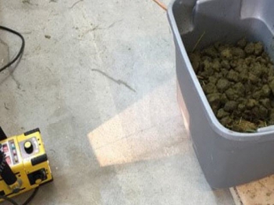
X-raying a bucket of manure to find the camera. University of Calgary Faculty of Veterinary Medicine photo.
Horses are notorious for gastric ailments. Over the course of a lifetime, chances are good that a horse will suffer at least once from some kind of gastrointestinal (GI) problem, with severity ranging from mild to life threatening, according to researchers with the University of Calgary in Alberta.
Because of the size of a horse, diagnosing GI issues can be challenging and require expensive specialized equipment.
“Horses are large animals, and the GI tract is a bit like a black box. It’s in the middle of the body, making it hard to see inside without being invasive,” said Dr. Renaud Léguillette, professor in the University of Calgary faculty of veterinary medicine (UCVM) and Calgary chair in equine sports medicine.
“You can use a long gastroscope, a 3 m-long soft tube with a camera at the end, but it only reaches the stomach, not the small intestine. You can use ultrasound, but the ultrasound doesn’t penetrate very deep, and the images don’t always show what you need to see,” he said.
However, a new tiny device — barely larger than a Tylenol gel cap — may make things easier for both horses and veterinarians.

Léguillette completed a clinical trial of a technique called capsule endoscopy, which involves a horse ingesting a "camera in a capsule." The camera travels through the animal’s GI tract and records video along the way. Similar technology has been used in human medicine for more than a decade, and Léguillette is pleased with the results in his equine patients.
“It’s an easy test to do. It’s not invasive, horses tolerate it very well and the images we get back are fantastic -- really exceptional,” Léguillette said. “It’s like a trip inside the small intestine. That’s really cool.”
The device records videos of the intestinal mucosa (mucous membrane that lines the GI tract) while the horse goes about its normal activities, the university explained. The system has four cameras, a LED light source and an onboard video recording system with a motion detection system.
“It gives you a 360-degree view of the inside of the GI tract,” Léguillette said.
Sedation or hospitalization isn’t needed. The veterinarian uses a nasogastric tube (a thin tube inserted up the horse’s nostril) to pump the camera through the esophagus and into the stomach.
“That’s the benefit of the technique. You don’t need special equipment. All you need is the camera and a nasogastric tube, which every large animal veterinarian has. You push the camera through, remove the tube and then it’s up to the horse to pass the camera in the manure and up to the owner to collect the camera,” Léguillette said.
Manure bins
While the procedure may be simple, recovering the camera is another matter, the researcher said. The time it takes the camera to transit the horse's GI tract varies from two to 10 days. During that time, all the horse's manure has to be collected and examined in order to recover the camera.
“It’s funny,” Léguillette said. “At first, we didn’t know how to do it, and my intern and summer student were screening the poop, washing it over a tub, and it was a big mess. It was a disaster, and of course, they were laughing.”
That’s when they came up with a better solution: collecting the manure in a big bin and then X-raying the bin every couple of days until the camera was found.
During the trial, Léguillette performed the capsule camera examinations on adult horses, foals, miniature donkeys and even a rhino, after getting a request from a U.S. zoo.
The results of the study showed that the capsule camera produces high-quality closeup images of ulcers, abrasions, polyps or small masses and bleeding and allows for a good examination of the stomach and small intestine, Léguillette said. Although it doesn’t work very well for the hindgut area that includes the colon, it offers another inexpensive tool in a veterinarian’s toolkit.
“For stomach and small intestine pathologies, this technique is excellent and readily available to veterinarians and horse owners,” Léguillette said.
Related news
 Best practices may reduce environmental mastitis cases
 Consequences of mastitis can be costly to a dairy, but treatment success varies depending on pathogen.
 Sodium butyrate may boost gain, feed efficiency in weaned heifers
 Supplementing the diets of post-weaned heifers with sodium butyrate may improve weight gain and feed efficiency, says researcher.
 Grass tetany preventable in grazing beef cows
 Plan ahead to minimize metabolic disorder by providing high-magnesium supplement prior to turning cows out onto lush spring pasture.