Vibriosis in Shrimp Aquaculture
Abstract
Aquaculture is the fastest growing food sector globally and is established itself as high protein resource to fulfill the food demand since the natural resources exhibits over exploitation. But, presently, the biggest problem faced by the aquaculture industry worldwide is diseases caused due to various biological and non-biological agents. Among the groups of microorganisms that cause serious losses in shrimp culture, the best known are bacteria because of the devastating economic effects they have on affected farms. Bacterial diseases, mainly due to Vibrio, have been reported in penaeid shrimp culture systems implicating at least 14 species and they are Vibrio harveyi, V. splendidus, V. parahaemolyticus, V. alginolyticus, V. anguillarum, V. vulnificus, V. campbelli, V. fischeri, V. damsella, V. pelagicus, V. orientalis, V. ordalii, V. mediterrani, V. logei etc
Vibriosis is one of the major disease problems in shellfish and finfish aquaculture. Vibriosis is a bacterial disease responsible for mortality of cultured shrimp worldwide (Lightner & Lewis, 1975; Adams, 1991; Lightner et al., 1992; LavillaPitogo et al., 1996; Lavilla-Pitogo et al., 1998; Chen et al., 2000). Vibrio species are widely distributed in culture facilitates throughout the world. Vibrio-related infections frequently occur in hatcheries, but epizootics also commonly occur in pond reared shrimp species. Vibriosis is caused by gram-negative bacteria in the family Vibrionaceae. Outbreaks may occur when environmental factors trigger the rapid multiplication of bacteria already tolerated at low levels within shrimp blood (Sizemore & Davis, 1985), or by bacterial penetration of host barriers. The exoskeleton provides an effective physical barrier to pathogens trying to penetrate the external surface of crustaceans, as well as the foregut and hindgut. However, Vibrio spp. are among the chitinoclastic bacteria associated with shell disease (Cook & Lofton 1973) and may enter through wounds in the exoskeleton or pores (Jiravanichpaisal & Miyazaki, 1994; Alday-Sanz et al,. 2002). The gills may appear susceptible to bacterial penetration because they are covered by a thin exoskeleton (Taylor & Taylor, 1992), but their surfaces are cleaned by the setobranchs (Bauer, 1998). The midgut, composed of the digestive gland (DG) and the midgut trunk (MGT, often referred to as the intestine, see Lovett & Felder, 1990), is not lined by an exoskeleton and therefore seems to be a likely site for penetration of pathogens carried in the water, food and sediment (Ruby et al., 1980; Jayabalan et al., 1982).
Vibrio harveyi, a gram-negative, luminous bacterium, is one of the important etiologic agents of mass mortalities of Penaeus monodon larval rearing systems. A large number of shrimp hatcheries (over 280) along the coastline of our country involved in shrimp seed production often suffer setbacks due to luminescent bacterial disease and suffer enormous economic losses.
Among the Vibrio harveyi isolates, some are virulent and some are not, suggesting a great deal of molecular and genetic variation in this group of bacteria. The pathogenic mechanism has also been recently attributed to bacteriophage.
Vibriosis is ubiquitous throughout the world and all marine crustaceans, including shrimps, are susceptible. Epizootics occur in all life stages, but are more common in hatcheries. Major epizootics of vibriosis have been reported for P. monodon from the Indo-Pacific region, P. japonicus from Japan, and P. vannamei from Ecuador, Peru, Colombia and Central America (Lightner, 1996). Vibriosis is expressed by way of number of syndromes. These include: oral and enteric vibriosis, appendage and cuticular vibriosis, localised vibriosis of wounds, shell disease, systemic vibriosis and septic hepatopancreatitis (Lightner, 1996).
Vibriosis is caused by a number of Vibrio species of bacteria, including: V. harveyi, V. vulnificus, V. parahaemolyticus, V. alginolyticus, V. penaeicida (Brock and Lightner, 1990; Ishimaru et al., 1995). There have been occasional reports of vibriosis caused by V. damsela, V. fluvialis and other undefined Vibrio species (Lightner, 1996).
Vibrio species are part of the natural microflora of wild and cultured shrimps (Sinderman, 1990) and become opportunistic pathogens when natural defence mechanisms are suppressed (Brock and Lightner, 1990). They are usually associated with multiple etiological agents. However, some Vibrio species, or strains of certain species, have been identified as primary pathogens (Owens and Hall-Mendelin, 1989; Owens et al, 1992; Lavilla-Pitogo et al., 1990; de la Peña a et al., 1995). Pathogenic strains of V. harveyi, V. vulnificus and V. parahaemolyticus have caused massive epidemics in Thailand (Nash et al., 1992) and the Philippines (Lavilla-Pitogo et al., 1990). Luminescent V. harveyi appears to release exotoxins (Liu et al., 1996) and may cause 80-100% mortality in P. monodon hatcheries (Harris, 1995). V. anguillarum, V. campbelli, V. nereis, V. cholerae (non 01) and V. splendidus have also been reported in association with disease outbreaks in shrimps (Chen 1992; Lavilla-Pitoga, 1990; Esteve & Quijada, 1993; Sahul-Hameed et al., 1996). The relationship between luminescence and toxicity of Vibrio carchariae in shrimp was investigated by Tatsuya Nakayama et al (2005). According to Jayasree. L. et al (2006) occurrence of five types of diseases: tail necrosis, shell disease, red disease, loose shell syndrome (LSS) and white gut disease (WGD) is by Vibrio spp. in Penaeus monodon from culture ponds of coastal Andhra Pradesh. Among these, LSS, WGD, and red disease caused mass mortalities in shrimp culture ponds. Six species of Vibrio—V. harveyi, V. parahaemolyticus, V. alginolyticus, V. anguillarum, V. vulnificus and V. splendidus—are associated with the diseased shrimp. The distribution and species composition of luminous bacteria in commercial penaeid shrimp hatcheries were studied by Jawahar Abraham. T and R. Palaniappan (2004). The observation on the presence of V. harveyi (97.30%) and V. orientalis (2.70%) in shrimp gut contents evinced that the primary source of these bacteria in a shrimp hatchery was the faecal matter from brood stock, possibly at the time of spawning.
Clinical signs
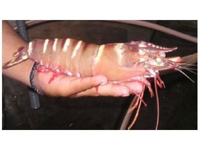
Mortalities due to vibriosis occur when shrimps are stressed by factors such as: poor water quality, crowding, high water temperature, low DO and low water exchange (Lewis, 1973; Lightner and Lewis, 1975; Brock and Lightner, 1990). High mortalities usually occur in postlarvae and young juvenile shrimps. P. monodon larvae suffered mortalities within 48 hr of immersion challenge with strains of V. harveyi and V. splendidus (Lavilla-Pitogo, et al., 1990). Mortalities involving vibriosis have been reported in market sized P. monodon (Anderson et al., 1988). Adult shrimps suffering vibriosis may appear hypoxic, show reddening of the body with red to brown gills, reduce feeding and may be observed swimming lethargically at the edges and surface of ponds (Anderson et al., 1988; Nash et al., 1992). Vibrio spp. also cause red-leg disease, characterised by red colouration of the pleopods, periopods and gills, in juvenile to adult shrimps and may cause mortality of up to 95% during the warm season (Chen, 1992). Eyeball necrosis diseases is caused by V. cholerae. The eyeballs of infected shrimps turn brown and fall away and mortality occurs within a few days (Chen, 1992). Six Vibrio species, including V. harveyi and V. splendidus cause luminescence, which is readily visible at night, in infected postlarvae, juveniles and adults (Ruby et al., 1980; Lightner, et al., 1992). Infected postlarvae may also exhibit reduced motility, reduced phototaxis and empty guts (Chen, 1992).
Gross Pathology
Shrimps suffering vibriosis may display localised lesions of the cuticle typical of bacterial shell disease, localised infections from puncture wounds, loss of limbs, cloudy musculature, localised infection of the gut or hepatopancreas and/or general septicemia (Lightner, 1993). Lesions of bacterial shell disease are brown or black and appear on the body cuticle, appendages or gills (Sinderman, 1990). Affected postlarvae may display cloudy hepatopancreata (Takahashi et al., 1985). Gills often appear brown (Anderson et al., 1988). Septic hepatopancreatitis is characterised by atrophy of the hepatopancreas with multifocal necrosis and haemocytic inflammation. Containing high loads of either Vibrio parahaemolyticus or V. harveyi induced the rounding up and detachment of epithelial cells from the basal lamina of the MGT. Epithelial cell detachment was not seen in the presence of non-pathogenic bacteria (probiotics) (Chen et al. 2000, Gary G. Martin et al. 2004). Pathogens like Vibrio spp., which cause detachment of the epithelium in the MGT, can affect high mortality in shrimp by eliminating 2 layers that protect the shrimp from infections: the epithelium and the peritrophic membrane it secretes. In addition, loss of the epithelium may affect the regulation of water and ion uptake into the body (Mykles 1977, Neufeld & Cameron 1994).
Histopathology
Systemic vibriosis typically results in the formation of septic haemocytic nodules in the lymphoid organ, heart and connective tissues of the gills, hepatopancreas, antennal gland, nerve cord, telson and muscle (Anderson et al., 1988; Mohney et al., 1991; Jiravanichpaisal et al., 1994). Infected hepatopancreocytes may appear poorly vacuolated, indicating low lipid and glycogen reserves (Anderson et al., 1988). Vibriosis in P. monodon is associated with the formation of “spheroids” in the lymphoid organ (Nash et al., 1992).
Diagnosis
Diagnosis of vibrio infection is based on clinical signs and the histological demonstration of rod-shaped Vibrio bacteria in lesions, nodules or haemolymph. Excised organs and haemolymph may be streaked on a Vibrio-selective (TCBS) or general marine agar plate. When investigating postlarvae, the whole animal may be crushed and then streaked onto an agar plate. Luminescent colonies may be observed after 12 to18 hr if incubated at room temperature or 25 to 30oC.
Vibrio isolates may be identified by a number of methods, including: Gram stain, motility, an oxidase test, mode of glucose utilisation, growth in the presence of NaCl, nitrate reduction and luminescence. Vibrio species may be identified rapidly in the field using the API-20 NFT system which involves culturing vibrio colonies on API-NFT strips and scoring the colonies according to the kit directions (Lightner, 1996) or BIOLOG (a miniaturised bacterial identification system which is an alternative to the API system). Antimicrobial sensitivity tests may be used to identify vibriosis and can be run using the Kirby-Bauer disk method (DIFCO, 1986) or the Minimum Inhibitory Concentration (MIC) method (Lightner, 1996)
Transmission Vibrio species exist in the water used in shrimp culture facilities (Lavilla-Pitogo, et al., 1990) and the biofilm, which is formed on different water contact structures of hatcheries and farms. Bacteria enter shrimps via wounds or cracks in the cuticle and are ingested with food (Paynter 1989; Lavilla-Pitogo et al., 1990). The primary source of V. harveyi in hatcheries appears to be the midgut contents of female broodstock, which are shed during spawning (Lavilla-Pitogo et al., 1992).
Viability
Numerous studies have been undertaken concerning the effect of freezing on vibrios which contaminate harvested shellfish. V. vulnificus in harvested oysters (Crassostrea virginica) survived storage at –20 oC for 70 days (Parker et al., 1994). V. parahaemolyticus, isolated from homogenates of oyster meat was inactivated within 16 days at –15 oC when the bacterial load was very high (10 cfu/gm; Muntada-Garriga et al., 1995). There is recent evidence to suggest that V. harveyi can survive in pond sediment even after chlorination or treatment with lime (Karunasagar et al., 1996).
Present status of vibriosis
Vibriosis is a common problem world-wide, particularly in India. V. harveyi continues to cause chronic mortalities of up to 30% among P. monodon larvae, postlarvae and adult under stressful conditions. A highly pathogenic strain of Vibrio sp. are also emerging and continues to cause mortalities among cultured shrimp (Le Groumellec et al., 1996). Problems caused by secondary vibriosis are common, but are considered minor compared to viral epidemics.
Treatment
Vibriosis is controlled by rigorous water management and sanitation to prevent the entry of vibrios in the culture water (Baticados, et al., 1990) and to reduce stress on the shrimps (Lightner, 1993). Good site selection, pond design and pond preparation are also important (Nash et al., 1992). An increase in daily water exchanges and a reduction in pond biomass by partial harvesting are recommended to reduce mortalities caused by vibriosis. Draining, drying and administering lime/dolomite to ponds following harvest is also recommended (Anderson et al., 1988).
Luminescent vibriosis may be controlled in the hatchery by washing eggs with iodine and formaldehyde and avoiding contamination by spawner faeces. V. harveyi in the water column can be inactivated by Chlorine Dioxide. Probiotics are administered directly into the water or via feeds. Immunostimulants have had success in reducing shrimp mortalities associated with vibriosis (Itami, 1996). Jiravanichpaisal and Chuaychuwong et al (1997) reported the use of Lactobacillus sp. as the probiotic bacteria in the giant tiger shrimp (P.monodon). They designed to investigate an effective treatment of Lactobacillus sp. against vibriosis and white spot diseases in P. monodon. They investigated the growth of some probiotic bacteria, and their survival in the 20 ppt sea water for at least 7 days. Inhibiting activity of two Lactobacillus sp. against Vibrio sp., E. coli, Staphylococcus sp. was determined.
The effect of copper concentration on the expression of both luminescence and toxin of V. harveyi was investigated by Nakayama. T. et al (2007). They found copper concentration of less than 40 ppm had no effect on the growth of shrimp. While V. harveyi cultured with 40 ppm copper concentration showed decreased luminescence. Therefore, the combination of prebiotics, probiotics, immunostimulants and non-antibiotic substances has superior specificity against vibriosis and Luminescent Bactria (LB) coupled with Best Aquaculture Practices (BAP), which makes it an effective management tool for the control of luminescence bacterial toxicity in aquaculture.
Dr A. Venkateswara Rao, Ph. D. Manager-Technical Services, Neospark Drugs and Chemicals Private limited Corporate Centre, 241, B.L. Bagh, Panjagutta, Hyderabad - 500 082, Andhra Pradesh, India
Related news
 Aquaculture Pond Cakes Trial Report
 Conducted product trials for Aquaculture Pond Cakes at Nellore, Telangana, India. The trial results shows - increased levels of Dissolved Oxygen