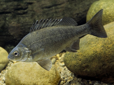
Diseases of the Australian Freshwater Fish Silver Perch (Bidyanus bidyanus) - Part 2
DIAGNOSTIC TOOLS
The following equipment is needed to perform on‑farm, disease diagnosis.
• Binocular compound microscope with powered light source, moveable stage, 10× ocular with 4×, 10×, 20× and 40× lens magnification (final magnification, 40×, 100×, 200× and 400×).
• Dissection kit including forceps, sharp‑pointed scissors, scalpel and blades, microscope slides and coverslips, cutting board and killing knife or pithing spike.
• Cast net, seine net and hand nets; buckets, airstones and airpump
Observation
Ponds, cages and tanks should be checked daily, in particular those with the following features.
• Recently (within 2 weeks) stocked or partially harvested;
• High stocking densities (particularly fingerling ponds);
• High feeding rates (>60 kg/ha; summer temperatures);
• Poor water quality or sudden variations e.g.
‘Crash’ of algal bloom
Low dissolved oxygen (DO) (<3 mg/L)
Very high temperatures (>30°C)
Sudden decreases in temperature (>4°C in 24 hrs)
High ammonia and high pH
Heavy surface scum, blue‑green algae blooms
Recent water exchange;
• Bird predation;
• Valuable broodfish.
Signs
Diseased fish display many physical and behavioural signs, and these can vary between diseases, levels of infestation or infection, size, age and condition of fish, culture facility and season. In general, any unusual signs in fish or unexpected changes in the water could be indicative of disease or pending disease. Some of the more common and important signs displayed by fish are:
• Loss of appetite – reduced feeding activity by some, many or all fish;
• Fish congregating near the surface, edges, aerators (Fig. 8);

Fig 8
• Fish swimming or acting abnormally, e.g. flashing;
• Fish with unusual colouring; may be pale or dark, eroded patches of skin, bulging eyes, red brains or enlarged and discoloured organs;
• Moribund or dead fish;
• Increasing numbers of moribund or dead fish.
• Presence or absence of feed in gut.
Water quality monitoring
The first action after the observation of unusual signs is to immediately monitor water quality with calibrated meters.
Ensure:
• DO (>3 mg/L), ammonia (<0.1 mg/L), pH (6 – 9.5) and temperature (10–30°C), these are minimum or maximum levels, or acceptable ranges. Management of suboptimal water quality
• Stop feeding;
• Increase aeration;
• Exchange water;
• Add lime to increase low pH (if <6.0 pH).
Sampling fish
Fish should be sampled (minimum 4 fish) (Fig. 9) after observation of the following conditions:

Fig. 9 and Fig. 10
• Mortalities and/or moribund (sick) fish;
• Sudden decrease in feeding behaviour (over 1–3 days);
• Abnormal swimming behaviour (‘flashing’; fish ‘listless’ and or ‘gasping’at pond edges or surface; fish positioned high in the water column or in water currents, circling or rapid swimming, rubbing on standpipes or ropes), abnormal colour;
• Fish unsighted for 2 weeks or more, particularly during autumn, summer and spring (>15°C water temperature).
Examination of gill and skin tissue
Live, preferably moribund fish collected for necropsy should be held in containers having aerated pond water (use water from pond). Microscopic examination should commence immediately following euthanasia (severing of spinal cord immediately behind head or pithing brain) as some parasites are prone to detaching and/or immobilisation soon after death of fish.
Using anaesthetics for euthanasia is not recommended as some ecto‑parasites may be killed or detach rapidly from the fish and will therefore be unseen during the examination. The following procedures are used to diagnose ecto‑parasitic and some fungal and bacterial diseases.
Gill examination – large fish:
• lift the opercular cover and using scissors, sever the white, cartilaginous gill arch (2 cuts) and remove >10 mm of gill (Figs. 10 and 11);

Figs 11,12,13
• place on a slide and using a scalpel, sever the cartilaginous gill arch from the primary gill lamellae (Fig. 12);
• discard the gill arch and add 2–3 drops of distilled water to the gill lamellae (do not use chlorinated tap water or pond water);
• lay a coverslip over the material and apply light pressure to encourage the lamellae to spread (Fig. 13);
• examine the tissue at low (40×), then higher magnification (100×).
Gill examination – small fish (<10 g):
• using scissors remove the opercular cover to reveal the gills;
• using scissors, sever a gill arch and remove a set of gills;
• separate one gill and discard the others; place the gill (cartilaginous gill arch attached) on a slide and add 2–3 drops of distilled water;
• lay a coverslip on the sample (the coverslip will be raised off the slide due to the thickness of the gill arch);
• add drops of water beneath the raised end of the coverslip until the tissue is ‘flooded’;
• examine the tissue.
Skin examination:
• position the caudal fin on a slide;
• using a scalpel and light pressure, scrape along the side of the body facing upwards (Fig. 14);

• remove skin/mucus from the scalpel to the slide;
• add 2–3 drops water, coverslip and examine as described for gill tissue.
Fin clip examination:
• remove a small piece of caudal fin (or any portion of fins that demonstrate abnormalities such as ragged edges) and prepare it as described for skin scraping.
Laboratory submission of fish for disease diagnosis When the disease cannot be diagnosed on‑farm, samples of fish should be delivered to a fish disease diagnostic laboratory. Moribund fish are the most suitable and reliable for diagnosis.
If delivery of live specimens is not possible, freshly euthanased specimens should be kept on wet ice and sent unfrozen provided they will arrive at the laboratory within 3 hours. If the delay is greater than 3 hours, the specimens should be sent preserved in 10% neutral buffered formalin. Tissue should not exceed 7 mm in thickness. In all cases the sample (organs or whole fish) should be representative of the population and ideally include moribund specimens.
Live specimens:
• package 4 to 6 fish in strong plastic bags; 1⁄₃ pond water, ²⁄₃ pure oxygen; air‑tight seal; watertight container;
• preferably overnight transport or deliver samples personally;
• do not feed fish prior to transport Iced specimens:
• wrap fish in a damp cloth or towel;
• place the sample in a plastic bag and cover with ice;
• place in a watertight container and deliver within 3 hrs.
Preserved specimens – small fish (<30 g):
• using scissors, open fish gut cavity from the vent to the gills (do not sever organs);
• place samples in a plastic bottle with 10% neutral buffered formalin;
• use 10 times the volume of fixative to tissue.
Preserved specimens – large fish:
• remove fat and dissect out organs (anterior and posterior kidney, gill, liver, heart, intestine, stomach, spleen, brain, skin with section of lateral line); sample any skin lesions ensuring a 5 mm margin of normal tissue is included; fix tissue/organs as for small fish.
Preserved specimens are required to be packaged in three watertight containers and labelled clearly prior to submission under IATA transport regulations. Check with your courier to ensure packaging meets recommended standards.
Frozen specimens:
• recommended for toxicological analysis and some forms of virology;
• frozen tissues are of little use for histopathology or microbiology (the two major diagnostic tools used in most fish disease investigations).
• freeze samples as soon as possible following the suspected poisoning.Information to accompany laboratory submissions:
• name; address; phone number and date
• species of fish; age and size
• water quality including pH; temperature; DO; ammonia; salinity
• size and type of production facility e.g. pond, cage, tank, raceway
• stocking density
• date mortality began and mortality rate per day
• clinical signs e.g. poor feeding; abnormal swimming; ‘flashing’
• water source
• diet; size and manufacturer; ration; Storage
• recent husbandry (past 2 weeks) e.g. harvesting; grading; water exchange; fish delivery
• system changes e.g. new tanks; changes in diet; chemical treatments
• environmental changes; weather conditions.
Laboratory Procedures
Specimens received at the laboratory will be processed and further examined.
• Histopathology – fixed or fresh tissues decalcified and individual tissues ‘cut in’ to assess single cell depth sections; stained; microscopically examined for pathogens and changes in tissue.
• Bacteriology – bacterial culture on specific media; targeted organs – kidney, spleen, brain, peritoneal fluids; also skin lesions; fresh tissue only (do not submit fixed tissue for bacterial culture); identification of bacteria; screening of bacterial colonies for antibiotic susceptibility (Fig. 15).

• Virology – viral culture on various fish cell lines; molecular diagnostics including PCR, sequencing and immunological tests to identify viruses.
Learning to recognise the ‘signs’ and diagnose disease Daily observation of fish, ponds and tanks, regular monitoring of water quality and a concise evaluation of skin and gill biopsies are the first steps used to investigate fish diseases. To the new or inexperienced fish farmer, interpreting physical signs and microscopic images is challenging.
However, the regular sampling of healthy and diseased fish will facilitate the recognition of normal and abnormal tissues, help in identification of the common pathogens, and develop the skills required to accurately diagnose disease. Most disease outbreaks in silver perch culture involve the common diseases and pathogens – during the early stages of an investigation look for the obvious!
Changes in the appearance of both skin and gill tissue are common during a disease event; however, determining the aetiological agent(s), or causes, is occasionally difficult due secondary invasion of pathogens or other organisms, or the manifestation of overt signs caused by systemic disease.
Changes in skin can include hyperaemia, haemorrhaging, ulceration, erosion, changes in pigment or thickening of the epithelium. Diseased gill tissue often shows hyperplasia and hypertrophy, causing cell growth and fusion between the secondary lamellae. Skin and gill tissue can have parasites and bacteria present without causing clinical disease; the interpretation of their significance will depend upon other clinical findings (Figs. 16, 17, 18, 19, 20, 21 and 22).


Maintaining water quality is critical in preventing disease; however, it is not always controllable. Water quality (DO, temperature, pH and TAN) should be monitored on a regular basis using highquality meters. Sudden changes in water quality e.g. an algae ‘crash’ (pond water turning tea colour), large temperature fluctuations (>5°C over 24 hrs), high TAN’s and periods of low DO (<3 mg/L) can cause stress and disease. Protozoan and fungal infections are particularly common, and observation of ponds should be daily following periods of poor or changing water quality.
Related news
 Improving the performance of cleaner fish in salmon aquaculture
 A new project aims to produce ballan wrasse that are more effective at eating sea lice on salmon farms.
 Food fit for a king (salmon)
 After seven years of research and development, a selection of advanced diets tailor-made for king salmon has finally been produced.
 Diseases of the Australian Freshwater Fish Silver Perch (Bidyanus bidyanus) - Part 1
 Diseases of the Australian Freshwater Fish Silver Perch (Bidyanus bidyanus) - Part 1